
The aforementioned reasons and the trend towards minimally invasive techniques have led to the increasing use of intraoperative 2D and 3D imaging and navigation in PS placement 9, 10, 11, 12.

However, the accuracy rates available in the literature differ substantially 8. The accuracy of freehand PS placement is generally considered acceptable. Another challenge is the interindividual differences in pedicle morphology, which require individual surgical planning before and possible adjustments during surgery 6, 7. Due to the close anatomical relation of the pedicle and sensitive structures, PS placement carries the risk of a number of complications, including neurological, vascular or dural injury following perforation of the pedicle wall 3, 4, 5. Pedicle screw (PS) placement has been considered a standard procedure in spine surgery for many years and is widely used for a variety of indications 1, 2. Weighted interrater reliability for Gertzbein Robbins grading was moderate for C-arm CBCT, substantial for CBCT and almost perfect for iCT. However, the exact reasons for the difference in accuracy remain unclear. Under quasi-identical conditions, higher screw accuracy was achieved with the combinations iCT/Curve and C-arm CBCT/Pulse compared with CBCT/StealthStation. Weighted interrater reliability was found to be 0.896 for iCT, 0.424 for C-arm CBCT and 0.709 for CBCT. Relevant perforations of the medial pedicle wall were only seen in the CBCT group. The differences between the different combinations were not statistically significant except for the comparison of C-arm CBCT/Pulse and CBCT/StealthStation (p = 0.003). Weighted kappa was used to calculate reliability between the observers. Grades A and B were considered acceptable and Grades C-E unacceptable. Two blinded observers classified screw placement according to the Gertzbein Robbins grading system. 470 screws were included in the final evaluation. With each combination of imaging system and navigation interface, 160 navigated screws were placed percutaneously in spine levels T11-S1 in ten artificial spine models. Thus, the objective of this study was to compare the accuracy of two combinations most used in the literature for spinal navigation and a recently approved combination of imaging device and navigation system. While different imaging and navigation devices can be used, there are few studies comparing these under similar conditions. Airo also connects seamlessly and automatically with Curve Information Guided Surgery by Brainlab, allowing for immediate surgical navigation on newly acquired images in the OR.3D-navigated pedicle screw placement is increasingly performed as the accuracy has been shown to be considerably higher compared to fluoroscopy-guidance.
#Brainlab airo portable#
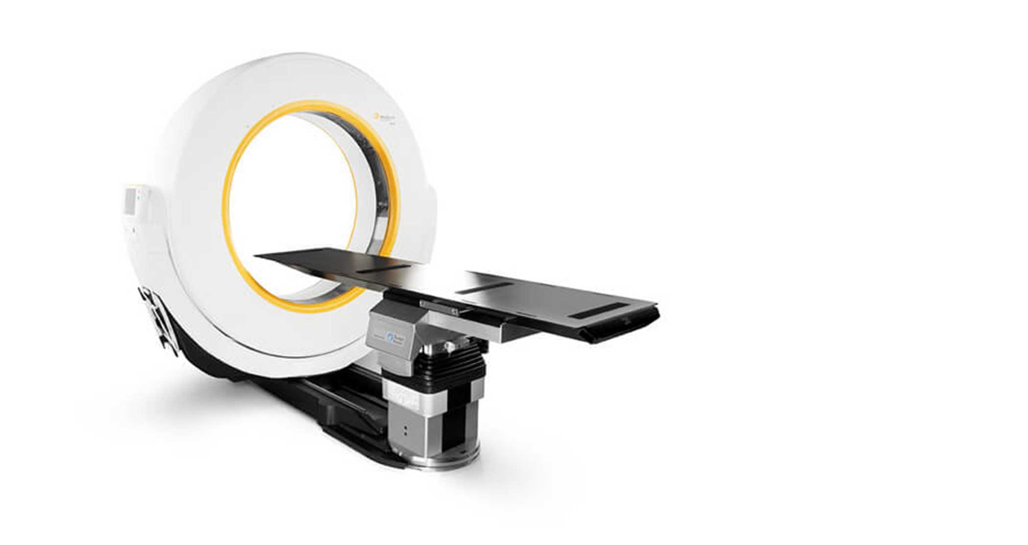
The portable scanner provides reproducible high-resolution images, thanks to its integrated OR table column by TRUMPF and 32-slice helical scan detector array.Īiro boasts the largest gantry opening on the market, lending itself particularly to cranial, spine and trauma procedures. Airo surpasses this demand and we are pleased to now be able to provide our European customers with this advanced imaging system.”Īiro is a unique system that advances healthcare delivery by producing high-quality CT images that enhance surgical decision-making and support minimally-invasive surgery. “We are dedicated to meeting the growing demand for real-time imaging to improve and expedite clinical decision-making which benefits both the patient and the surgical team.


“In light of the enormous market response Airo has already experienced around the world, we are eager to begin installation and use in Europe,” said Stefan Vilsmeier, president and chief executive at Brainlab. The CE Mark allows Brainlab to begin installation of the systems sold in the European Union. Global sales and CE Mark approval represent significant milestones for innovative systemīrainlab and Mobius Imaging have announced CE Mark approval for the Airo Mobile Intraoperative CT.


 0 kommentar(er)
